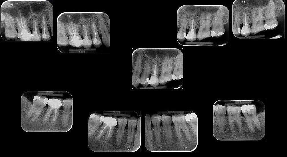
Periapical radiography is an intraoral technique for examining a single tooth and surrounding tissues. Each film represents the features and details of the tooth and alveolar bone. The most important clinical features in periapical radiography including:
- Evaluation and diagnosis of dental caries
- Assess the condition of the tissue around the tooth
- Evaluation of the condition of the tooth and its surrounding alveolar bone after trauma
- Assess the position of the impacted teeth
- Evaluation of tooth root morphology and its channels
- Evaluation before and after apical surgery
- Accurate evaluation of apical cysts and other lesions associated with alveolar bone
- Assess the condition of the implant
- Evaluation of infection or inflammation of the apical dental section
In order to take accurate photos, it is very important how the film, the holder and the radiation source are placed. Because the anatomy of the oral cavity is different among people, to achieve better results, two techniques are used in periapical imaging, which are:
- Parallel technique
- Bisect technique
Due to the fact that the parallel technique is ideal due to the geometry of the film, teeth and X-rays, in the Dr. Safi’s center, the parallel technique is always used.